
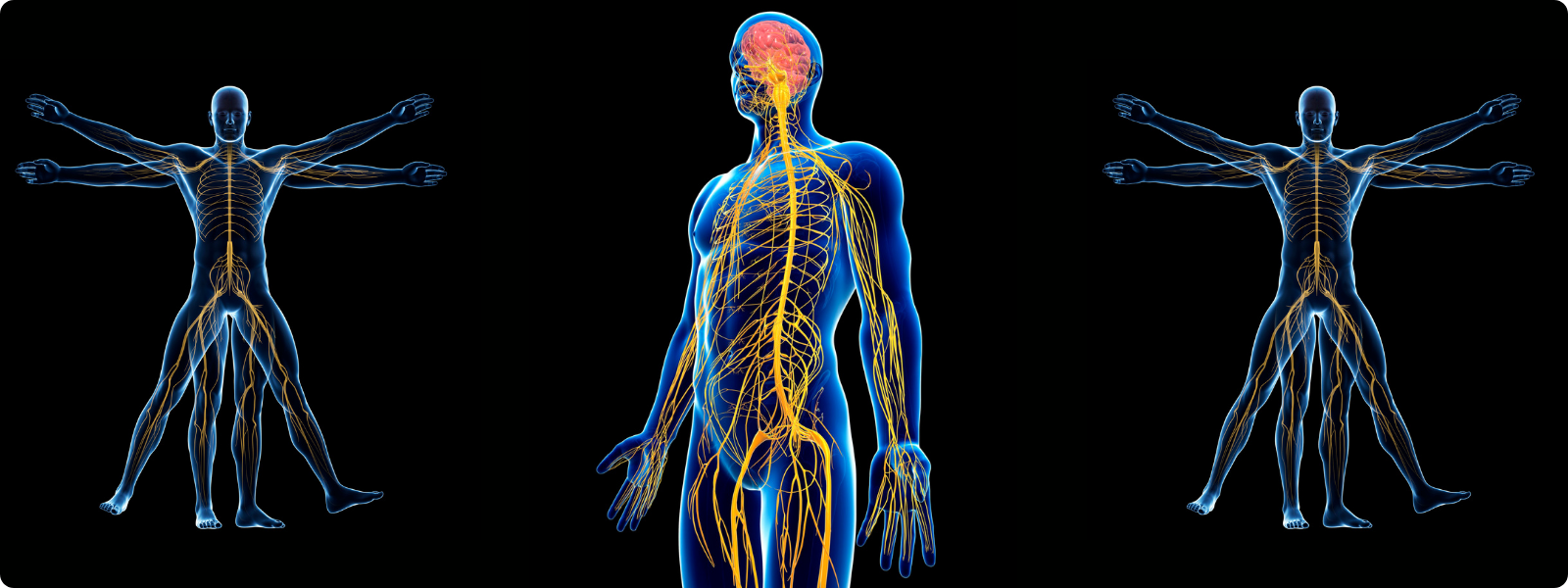
Spine To Nerve Connection
A widespread disconnect still exists, even within healthcare regulatory bodies, about chiropractic's impact on nervous system function. In this article, we review some of the most interesting and relevant literature connecting the spinal system to the nervous system to build a literature-based foundation for communicating with patients, the general public, medical providers, politicians, and regulatory figures.
Anatomy
Your spinal system is a complex network of vertebrae (i.e., bones), discs (i.e., thick cartilage between the bones), ligaments (i.e., connective tissues that surround every aspect of the spinal bones and discs), arteries (i.e., running along the sides of the spine and directly in front of your spine), cerebrospinal fluid (i.e., liquid inside the spine that protects nerves and supports nutrition and waste pathways), and nerves (the central nervous system is inside your spine; spinal nerve roots and vagus nerves outside of your spine).
From a nerve perspective, there are two parts of your central nervous system which are located inside of your spine: the lower part of your brainstem is inside of C1, and the entirety of your spinal cord is located inside C2 (the second bone of the spine) through L1-L2 (the first and/or second bone of the lumbar spine). Your central nervous system is important because it regulates all other systems, including how tissues repair and regenerate after physical or chemical trauma. (2)
Additionally, the vagus nerve (cranial nerve X) exits the inside of the spine at the brainstem level, above the level of C1, and runs directly in front of C1, posterior to the carotid sheath, as they travel down the front of the spine, connecting the brain with the heart and many organs. (7, 8) Of equal importance are the C1 nerves (being the nerve root located between the C1 and C2 vertebrae of the spine), which have been described as the body’s EMT system, responsible for blood pressure as well as “physiological processes ranging from neuroendocrine responses to infection and inflammation, glucose homeostasis, reproduction, breathing, thermoregulation, hypothalamic-pituitary axis (HPA) -mediated stress responses, and food consumption”. (13) These studies show that cardiac pain, organ referral pain (which can commonly present as back, neck and/or jaw pain) and angina is most often vagus nerve dysfunction that is transmitted to the brain through the C1-C2 spinal segments. (4. 9) Another fascinating study shows that the C1 nerves mediate inflammation directly through altering the function of the immune system both within state of pathology such as kidney disease and within normal homeostasis and physiology. (5, 6) The critical importance of the nervous system to human function and adaptability is likely why the notochord (which becomes the vertebral discs and spinal joints) and neural ectoderm (which becomes the brain, spinal cord and peripheral nerves) are considered the first tissue-structures to form in embryonic development. (1, 2)
Your spinal system is also designed to support human movement; flexion, extension, lateral movement and twisting which adds complexity to the spinal system and how it is connected with the nerves inside and outside of it, which does not exist in the skull-brain connection as there is limited movement in that part of human anatomy. Spinal movement is also more than just convenient for life. Spinal movement leads to electrical nerve signals (mechanoreception and proprioception) that are constantly being sent from the spinal cord to the brain for the purpose of letting the brain know where the spine is in space as well as help keep the brain functioning normally and tissues healthy. This is one of the reasons why when a person has a sedentary lifestyle with limited or no movement, their brain and their spine will become abnormal and dysfunctional.
Damage to the spinal system, such as ligament instability, misalignment that compromises normal spinal movement or causes neural-arterial compression, and/or abnormal inflammation caused by an unhealthy spinal system, can negatively affect normal nerve function. (3, 27, 28)
Chiropractic Literature
Now that you understand some of the basics of spinal anatomy, it will be easier to see how chiropractic care might improve neurological function, with a myriad of health benefits as a byproduct.
A 2015 study in the Journal of Experimental Brain Research showed that a single chiropractic treatment improved cortical drive (measured via SEMG on the maximum voluntary contraction of planar flexors) and reduced muscle fatigue compared to the control group. (10)
A 2016 study published in the Journal of Manipulative and Physiological Therapeutics showed improvement in sensorimotor function and multiple sensory integration in elderly adults (through testing static posture, ankle joint position sense, sound-induced flash illusion and choice stepping time) following three chiropractic adjustments. (15) Sensorimotor function is a function of the central nervous system. (16)
A 2016 study also published in the Journal of Manipulative and Physiological Therapeutics showed very interesting findings in a cohort of women, some who were in their second trimester of pregnancy and some who were not pregnant. The women who received chiropractic care while pregnant showed an increase in elevator hiatal area indicating relaxation of the pelvic floor which was confirmed with trans-perineal ultrasound. This relaxation of the pelvic floor muscles did not occur in the non-pregnant cohort which indicates that chiropractic care is not only changing how the brain regulates muscles, but that the change is intelligent to specific life stages. (17) This critical understanding of chiropractic care which works with the body towards normal function, differs greatly from pharmacology which is often dose dependent towards a particular outcome regardless of whether that outcome is helpful or harmful to the body.
A 2016 study published in the journal of Neural Plasticity showed that a single chiropractic adjustment improved somatosensory processing at the cortical level, specifically within the prefrontal cortex as verified on 62-lead brain EEG. (19) This study shows untapped potential for chiropractic care in the management and reversal of mental health problems as the prefrontal cortex is one of the primary centers of the brain responsible for emotional regulation, decision making, social cognition, and psychopathology. (20)
A 2018 study published in the Journal of Electromyography and Kinesiology showed that a single chiropractic treatment reduces the cortical silent period and increases the threshold for motoneurons excitability in the low limb muscles. (11) What this means in lay terms is that a single chiropractic adjustment is showing a positive effect on the brain’s ability to regulate leg muscle function. Additionally, this non-chiropractic study published in the medical journal Clinical Neurophysiology in 2020 reports that the ability to modulate motoneuron excitability is “profoundly important for the clinical diagnosis and treatment of neurological disorders”. (13)
A 2019 meta-analysis published in the journal Headaches found that multiple studies indicated chiropractic treatments reduced migraine days, pain, and intensity. (18)
A 2020 study published in ACTA Biomedica indicates that chiropractic care and adjustments help regulate the autonomic nervous system at both the peripheral and central nervous system levels and could be considered an “effective therapy for major depression”. (21)
Several studies published in 2019 and 2020 in the journals Brain Science and Scientific Reports indicate that a single session of chiropractic care increased the amplitude of the N30 SEP peak (i.e., brain function) as well as the patient’s functional connectivity between different parts of the brain in chronic stroke patients, as verified by brain EEG. (22, 23) Additionally, increases in muscle strength were associated with increases in the V-wave and H-reflex, further indicating that chiropractic care changes central nervous system function and regulation. (24)
A 2021 study published in the European Journal of Pain discussed that neck and back pain are the primary causes of disability worldwide, accounting for more than 10% of the total years lived with disability. The study reviews the mechanisms of chiropractic care and the likelihood that chiropractic care inhibits pain by changing spinal cord function. (14)
A 2024 study published in the journal of Brain Science showed that changes in brain waves following chiropractic care led to improved overall quality of life, including significant improvement in sleep as well as reduction in anxiety, depression, fatigue and pain. (25)
A 2024 study published in the medical journal Cureus discusses the “multifaceted influence of chiropractic care on processes of the neuroendocrine-immune system and integrated physiology,” including functional improvements in blood pressure, heart rate variability, and sleep following chiropractic care to the spinal system. (26)
In conclusion, published scientific evidence suggests that chiropractic adjustments positively affect nervous system function through treatment of the spinal system. Hopefully, this data can become regular discussion points within chiropractic offices so that someday in the near future, the spine-to-nerve connection can become a basic understanding for the general public, medical providers, politicians, and regulatory figures.
(1) Elshazzly, M., Lopez, M. J., Reddy, V., [add remaining authors]. (2023). Embryology, central nervous system. In StatPearls. StatPearls Publishing. https://www.ncbi.nlm.nih.gov/books/NBK526024/
(2) Akinrodoye, M. A., & Lui, F. (2022). Neuroanatomy, somatic nervous system. In StatPearls. StatPearls Publishing.
(3) Lucas Lucio, L., & de Andrade Lourenção Freddi, T. (2023). Glossopharyngeal, vagus and accessory nerves: Anatomy and pathology. Seminars in Ultrasound, CT and MRI, 44(2), 95–103. https://doi.org/10.1053/j.sult.2022.11.003
(4) Foreman, R. D., Garrett, K. M., & Blair, R. W. (2015). Mechanisms of cardiac pain. Comprehensive Physiology, 5(2), 929–960. https://doi.org/10.1002/cphy.c140032
(5) Inoue, T. (2021). Neuroimmune system-mediated renal protection mechanisms. Clinical and Experimental Nephrology, 25(9), 915–924. https://doi.org/10.1007/s10157-021-02062-3
(6) Abe, C., & Inoue, T. (2018). Role of C1 neurons in anti-inflammatory reflex: Mediation between afferents and efferents. Neuroscience Research, 136, 6–12. https://doi.org/10.1016/j.neures.2018.05.001
(7) Dionigi, G., Chiang, F. Y., Rausei, S., Wu, C. W., Boni, L., Lee, K. W., Rovera, F., Cantone, G., & Bacuzzi, A. (2010). Surgical anatomy and neurophysiology of the vagus nerve (VN) for standardised intraoperative neuromonitoring (IONM) of the inferior laryngeal nerve (ILN) during thyroidectomy. Langenbeck’s Archives of Surgery, 395(7), 893–899. https://doi.org/10.1007/s00423-010-0693-3
(8) Kronsteiner, B., Carrero-Rojas, G., Reissig, L. F., Moghaddam, A. S., Schwendt, K. M., Gerges, S., Maierhofer, U., Aszmann, O. C., Pastor, A. M., Kiss, A., Podesser, B. K., Birkfellner, W., Moscato, F., Blumer, R., & Weninger, W. J. (2024). Characterization, number, and spatial organization of nerve fibers in the human cervical vagus nerve and its superior cardiac branch. Brain Stimulation, 17(3), 510–524. https://doi.org/10.1016/j.brs.2024.04.016
(9) Myers, D. E. (2021). The receptive field for visceral pain referred orofacially by the vagus nerves. Clinical Anatomy, 34(1), 24–29. https://doi.org/10.1002/ca.23604
(10) Niazi, I. K., Türker, K. S., Flavel, S., Kinget, M., Duehr, J., & Haavik, H. (2015). Changes in H-reflex and V-waves following spinal manipulation. Experimental Brain Research, 233(4), 1165–1173. https://doi.org/10.1007/s00221-014-4193-5
(11) Haavik, H., Niazi, I. K., Jochumsen, M., Uginčius, P., Sebik, O., Yılmaz, G., Navid, M. S., Özyurt, M. G., & Türker, K. S. (2018). Chiropractic spinal manipulation alters TMS induced I-wave excitability and shortens the cortical silent period. Journal of Electromyography and Kinesiology, 42, 24–35. https://doi.org/10.1016/j.jelekin.2018.06.010
(12) Heckman, C. J., Mottram, C., Quinlan, K., Theiss, R., & Schuster, J. (2009). Motoneuron excitability: The importance of neuromodulatory inputs. Clinical Neurophysiology, 120(12), 2040–2054. https://doi.org/10.1016/j.clinph.2009.08.009
(13) Guyenet, P. G., Stornetta, R. L., Bochorishvili, G., DePuy, S. D., Burke, P. G., & Abbott, S. B. (2013). C1 neurons: The body’s EMTs. American Journal of Physiology-Regulatory, Integrative and Comparative Physiology, 305(3), R187–R204. https://doi.org/10.1152/ajpregu.00054.2013
(14) Gevers-Montoro, C., Provencher, B., Descarreaux, M., Ortega de Mues, A., & Piché, M. (2021). Neurophysiological mechanisms of chiropractic spinal manipulation for spine pain. European Journal of Pain, 25(7), 1429–1448. https://doi.org/10.1002/ejp.1773
(15) Holt, K. R., Haavik, H., Lee, A. C., Murphy, B., & Elley, C. R. (2016). Effectiveness of chiropractic care to improve sensorimotor function associated with falls risk in older people: A randomized controlled trial. Journal of Manipulative and Physiological Therapeutics, 39(4), 267–278. https://doi.org/10.1016/j.jmpt.2016.02.003
(16) Murray, G. M., & Sessle, B. J. (2024). Pain-sensorimotor interactions: New perspectives and a new model. Neurobiology of Pain, 15, Article 100150. https://doi.org/10.1016/j.ynpai.2024.100150
(17) Haavik, H., Murphy, B. A., & Kruger, J. (2016). Effect of spinal manipulation on pelvic floor functional changes in pregnant and nonpregnant women: A preliminary study. Journal of Manipulative and Physiological Therapeutics, 39(5), 339–347. https://doi.org/10.1016/j.jmpt.2016.04.004
(18) Rist, P. M., Hernandez, A., Bernstein, C., Kowalski, M., Osypiuk, K., Vining, R., Long, C. R., Goertz, C., Song, R., & Wayne, P. M. (2019). The impact of spinal manipulation on migraine pain and disability: A systematic review and meta-analysis. Headache, 59(4), 532–542. https://doi.org/10.1111/head.13501
(19) Lelic, D., Niazi, I. K., Holt, K., Jochumsen, M., Dremstrup, K., Yielder, P., Murphy, B., Drewes, A. M., & Haavik, H. (2016). Manipulation of dysfunctional spinal joints affects sensorimotor integration in the prefrontal cortex: A brain source localization study. Neural Plasticity, 2016, Article 3704964. https://doi.org/10.1155/2016/3704964
(20) Hiser, J., & Koeni